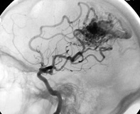
- Patient with brain AVM involving the left side of the brain.
- Pre-radiosurgery embolization was performed to decrease the size of the AVM.
- Angiogram at left shows abnormal tangle of blood vessels following injection of the left internal carotid artery with x-ray dye.
- The patient is facing to the left in this image.

Treatments & Services
Arteriovenous Malformation Embolization
The patient was brought to the angiography suite and placed under general anesthesia. A catheter was placed through the patient's groin upstream of the brain AVM. Tiny catheters were then systematically advanced under x-ray guidance into the larger vessels feeding the abnormal tangle of vessels. In this case, a special medical grade liquid adhesive was carefully injected under x-ray guidance through the tiny catheters into the AVM. This resulted in decreased flow through the AVM. Below are images of the glue that was injected into the AVM.
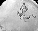
The image to the left shows the glue that was deposited into the AVM from the same side view. You can see the effect of this glue on the next two images which compare the AVM before and after treatment.
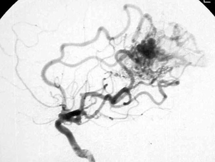
Before Treatment
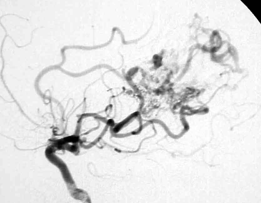
After Treatment
There is significantly decreased blood flow through the AVM following embolization with the liquid adhesive. This patient went on to have radiosurgery for the remaining AVM.